


VisiScope Cell Explorer


VisiScope Cell Explorer
Life Cell Imaging System
The new challenge in Digital Fluorescence Microscopy: Multidimensional Acquisition and Analysis of living CellsModern cell biology on living cells and tissues has reached revolutionary success in biological, medical and pharmacological research in recent years. With the VisiScope system you can reliably document living organisms and intracellular processes, even over a period of several days. While someone has to work with none-automated microscopes resulting in restricted application possibilities in the past, nowadays fully automated fluorescence microscopes with multidimensional control have been established. The VisiScope Cell Explorer, based on Visitron imaging software, solves all these new challenges easily and user friendly with highest precision.
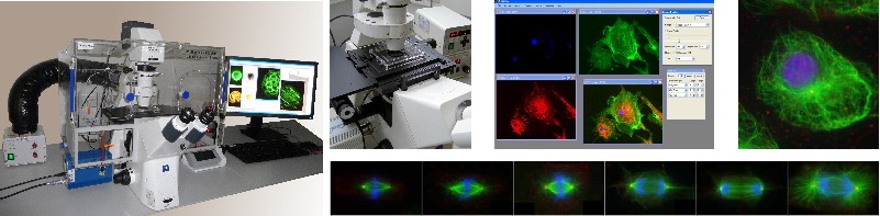 |
VisiScope Cell Explorer – Overview of System Components
- High-performance microscope with superior optics and automation
- High resolution digital scientific camera with highest sensitivity and best cooling
- Fully automated xy-table with best position accuracy and reproducibility
- Precise z-axis control and acquisition (optionally: piezo z-focus drive)
- Powerful Visitron imaging software including multidimensional control of all hardware / motorized components
- Incubation chambers controlling cell culture environment
Click here to find out more